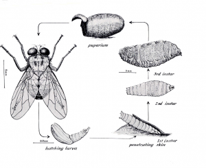
NICD has been consulted about numerous cutaneous maggot infestations (furuncular myiasis) in humans in North West Province, as well as increased sporadic cases in Gauteng Province. Our laboratory has confirmed that the maggots are those of the tumbu fly (also known as the ‘mango’ or ‘putsi’ fly). The increase in the number of cases is most likely related to the recent marked increase in seasonal rainfall, leading to the expansion of the fly’s normal range, namely the warmer northern and eastern parts of the country. The adult female tumbu fly (Cordylobia anthropophaga) deposits eggs usually on urine- or faeces-contaminated sand, soil or clothing. The larvae (maggots) hatch and on contact with skin, penetrate and cause enlarging boil-like skin lesions, each with a small opening at the apex through which the larva breathes (furuncular myiasis)(Figure). The lesions may be complicated by secondary bacterial infection. The condition is readily treated by applying petroleum jelly (Vaseline) or liquid paraffin to the lesions, to suffocate the maggots and lubricate the cavity in the skin; usually, they then emerge or are easily expressed with finger pressure. Incision or use of forceps or other instruments is unnecessary and should be avoided, as inflammation or secondary infection is more likely if the larva and/or skin is damaged. Domestic dogs and rodents are commonly affected, sometimes with large numbers of lesions.
Prevention: washing should not be laid on the ground to dry. Ironing of clothes will kill eggs or larvae. Affected dogs should be dipped in an appropriate insecticide solution, as for prevention of tick or flea infestation, under veterinary guidance.

Figure. The life cycle of Cordylobia anthropophaga. Larval instars 1-3 are the stages causing the skin lesions. From: Zumpt F. The Arthropod Parasites of Vertebrates in Africa South of the Sahara. Vol III. Publ. of the SAIMR (1966); 13(52): 48-49.


